...Julie Newdoll merges life science and culture, myths and molecules in her
paintings, music, journal covers and science games.

Oil Painting, 20 x 20, 2015, for Molecular and Cellular Proteomics Poster.
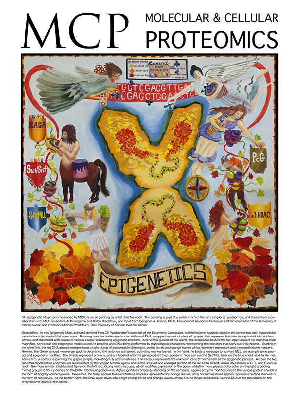
“An Epigenetic Map”, commissioned by MCP, is an oil painting by artist Julie Newdoll. This painting is part of a series in which the artist explores proteomics, and stems from a collaboration with MCP co-editors Al Burlingame and Ralph Bradshaw, and input from Benjamin A. Garcia, Ph.D., Presidential Associate Professor and Simone Sidoli of the University of Pennsylvania; and Professor Michael Washburn, The University of Kansas Medical Center. Description: In this Epigenetic Map, a phrase derived from CH Waddington’s concept of the Epigenetic Landscape, a chromosome-shaped island in the center has both inaccessible mountainous terrain and flat open areas. Running over the landscape is a red ribbon of DNA, wrapped around clusters of grapes that represent histones incorporated into nucleosomes, and decorated with leaves of various colors representing epigenetic markers. Around the outside of the island, the accessible DNA of the flat, open area of the map has been magnified, so we can see epigenetic modifications to proteins and DNA being performed by mythological characters representing the enzymes that carry out this process. Starting in the lower left, the red DNA strand emerges from a tight clump of inaccessible chromatin, buried in red and orange leaves which represent repressive and transient histone markers. Hermes, the Greek winged messenger god, is decorating the histones with green, activating marker leaves. In his hand, he holds a message to activate MLL, an example gene product and epigenetic modifier. The shields represent proteins, and are labelled with the gene product they represent. You can see the Set/MLL label on the blue shield next to Hermes. Above him, a centaur is painting the grapes purple, indicating fully active histones. The centaur represents the unknown control mechanism of the epigenetic process. Across the top, two DNA modification enzymes are represented by the winged female figures above the unfurled and enlarged portion of the red DNA strand, where DNA bases A, G, T, and C can be read. The more sinister, bird-bodied figure on the left is oxidizing methyl groups, which modifies expression of the gene, while the more pleasant character on the right is adding methyl groups to the cytosines on the DNA. Continuing clockwise, Aglaia, goddess of beauty standing on the compass, applies enzyme modifications to the various protein shields in the form of brightly colored jewels. Below her, the Satyr applies transient histone markers represented by orange leaves, while his female muse applies repressive histone markers in the form of red leaves. At the bottom right, the DNA again dives into a tight clump of red and orange leaves, where it is no longer accessible, like the DNA in the mountains on the chromosome island in the center. .
Computer Graphics, hand drawing and digital collage, 18 x 24, internal project Gilead, 2014
An example of work done for product release and other promotions, involving a combination of hand drawn artwork and digital, by Julie Newdoll.

Mixed Media on Plate, cover of MCP Proteomics April 2013; 12 (4).
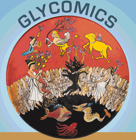
On the cover: (Oil painting by Julie Newdoll, www.brushwithscience.com): A cell membrane constructed of the half human, half fish (both water loving and land creature) Oannes of Mesopotamian mythology divides the image into two worlds - that of the cytoplasm (black) below the outside the cell (red) above. In the cytoplasm reside the water creatures - species of fish and sea monster. They are glycosylated, if at all, with a single sugar GlcNAc, represented by the blue and white dragonfly (in the larvae stage in the water and as a dragonfly on the outside of the cell). Spanning the membrane are various characters from Greek mythology representing membrane proteins. They all hold or are attached to a straight or branched object, which represent straight or branched sugars. At the end of the chain of sugars is a purple bird, representing Neu5Ac. The objects in these glycans, like the bird, are colored to reflect the “Recommended symbols and conventions for drawing glycan structures” published in the Essentials of Glycobiology*. Hera (or Juno) holds her golden apple tree full of Gal, GalNac, and GalN. Cerberus sports a snake on each head, which might be a short chain of IdoA and GalA, all with the purple bird on the end of each branch (Neu5Ac), while the ram with the golden fleece has only a single GlcNAc, reflecting the possibility that proteins in the cytoplasm may be glycosylated in this way as shown recently in the literature. For details, see Essentials of Glycobiology, 2009, 2nd edition, Varki A, Cummings RD, J.D. Esko, et al., eds., Cold Spring Harbor Laboratory Press, Cold Spring Harbor, NY.
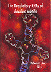
Painting on the cover of The Regulatory RNAs of Bacillus subtilis, thesis by Ruben Mars, Ph.D., Univeristy of Gronigen, 2014
RNA is best known for its role in protein synthesis where messenger RNA (mRNA) directs protein synthesis on ribosomes. However, it is becoming increasingly clear that many RNA molecules have regulatory rather than protein-encoding functions. Cover art by Julie Newdoll.

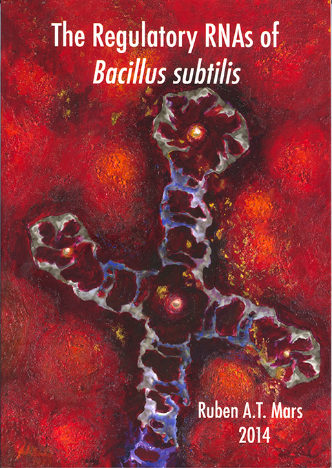
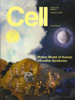
Digital collage of watercolor artwork for Cover of Cell July 3, 2013, Vol. 154. No. 1 pp. 1-252.
On the cover: Developmental mechanisms generate interspecies hybrids that cannot propagate, as alluded to by the artist's depiction of the mythical Minotaur's labyrinth on the cover. However, even socially naïve animals rarely mate with members of other species. The mechanisms that control such behavioral reproductive isolation, an important tenet of evolution, are not well understood. As depicted on the cover, male flies tap other flies with their foreleg prior to attempting to mate. Fan et al. (pp. 89–102) show that foreleg removal permits males to mate with flies of other species. Activation of foreleg neurons expressing the chemoreceptor Gr32a is necessary and sufficient to inhibit interspecies courtship by D. melanogastermales. Gr32a is required to detect aversive hydrocarbons found on the cuticle of other drosophilid species, and one such hydrocarbon, z-11-pentacosene, is visible on the female fly on the cover. Strikingly, female flies utilize a Gr32a-independent mechanism to reject males of other species, with the cover depicting the female fly rejecting the male who is tapping her. Cover art by Julie Newdoll.
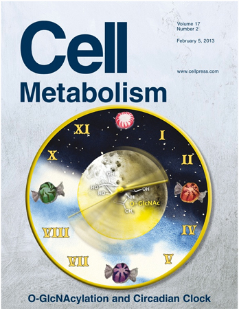
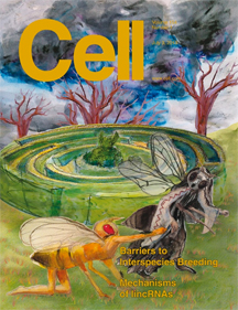
Digital collage of watercolor artwork for Cell Metabolism, Feb. 25, 2013, Vol. 17, No.2
On the cover: The circadian clock plays an important regulatory role in a wide range of metabolic functions. On pp. 303–310 and 291–302 of this issue, two studies by Li and colleagues and Kaasik and colleagues show that the hexosamine/O-GlcNAc pathway acts as a glucose sensor to modulate circadian clock oscillations. While O-GlcNAcylation opposes the effects of ubiquitination to stabilize clock proteins, it also exhibits a reciprocal regulation with GSK3β-mediated phosphorylation to fine tune the circadian clock. The cover image illustrates how clock speed is regulated by glucose levels (candies) sensed via O-GlcNAcylation, as shown by Li and colleagues. Cover art by Julie Newdoll.
On the cover: Proteasome Bashe Dragons Devouring and Digesting Ubiquinated Substrates. Watercolor painting by Julie Newdoll, 12 x 18, 2012, sold.
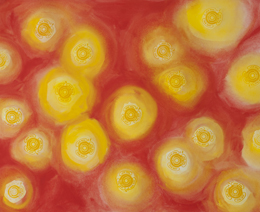
In the cytosol (the ocean region in the painting) certain protein “fish” have been tagged for degradation by a seaweed-like chain of ubiquitin wrapped around their tails. One such fish is being shuttled towards the proteasome “dragon” by a fish-tailed sea god, while others are drifting towards destruction on their own. After digestion, the protein “fish” can be seen expelled from the other end of the dragon as bones. In Chinese mythology, Bashe Dragons are said to eat elephants and excrete their bones years later. In the nucleus, the bright orange land of the underworld in the painting, the black silhouette of a second proteosome dragon can been seen involved in the same task. This dragon is similar, but not the same, as the one in the cytosol in ways that are not yet understood.


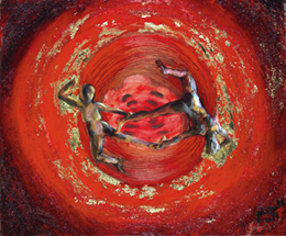
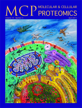
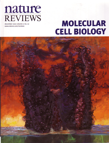
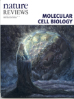
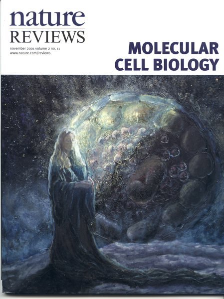
“The Worlds of Proteomics”, oil on canvas, 33" x 44", $4,000. The painting is the first of a series in which the artist will explore proteomics, and stems from a collaboration with MCP co-editors Al Burlingame and Ralph Bradshaw. The following article appeared in ASBMB News
The Beauty of Proteomics
BY ANGELA HVITVED
It may be beautiful, but the April cover of Molecular and Cellular Proteomics is not just another pretty picture. The cover artwork, commissioned by MCP, is an oil painting by artist Julie Newdoll titled “The Worlds of Proteomics.” The painting is the first of a series in which the artist explores proteomics and stems from a collaboration with MCP co-editors Al Burlingame and Ralph Bradshaw. Newdoll worked with Burlingame and Bradshaw to learn about the many aspects of proteomics research and to understand the broad scope of science that MCP publishes. Their goal in initiating the series is to promote a deeper, richer appreciation for the field by those directly and indirectly involved with proteomics research.
Newdoll has a master’s degree in medical illustration and previously worked as a visualization specialist at the University of California, San Francisco. She found that fusing the powerful and beautiful concepts of science with cultural references both ancient and contemporary provided a new lens for creating her own view of the fascinating world of science. Other commissioned paintings by Newdoll can be found at her website.
The painting on the cover is accompanied by the artist’s description, which explains Newdoll’s inspiration for the piece:
“When proteins are made in the cell in response to some stimuli or event, they are targeted via an address system for a specific location or locations. In this painting, the various areas in a cell are represented by various worlds – there is the world of the sea in the cytosol, that of the air outside the cell and land or earth inside the nucleus. Inside the mitochondria and the endoplasmic reticulum, little islands have their own color scheme. A protein meant to be secreted to the outside of the cell follows an elaborate path of production, first forming inside the endoplasmic reticulum, later packaged into a membrane bubble which melds with the Golgi, and finally repackaged and released to the outside of the cell.
“From the artist’s perspective, a protein meant for the outside of the cell is rendered as a flying creature in this painting. It never ends up in the deep sea environment of the cytosol, or it would ‘drown.’ Likewise, proteins destined for the deep sea of the cytosol could not breathe outside the cell in the open air. And then there are the amphibians …”
“The Worlds of Proteomics” represents the broadest perspective of proteomics research, and Newdoll intends to use it as the foundation of the series. “The first in the series displays all realms of proteomics, while future paintings will zoom in on the various worlds and microcosms mapped out here,” she explains. The co-editors intend to feature future pieces on the journal’s cover to highlight specific areas of research.
To celebrate the debut of the new cover artwork, MCP, normally an online-only publication, has printed 500 copies of the April issue as well as a poster of the painting for distribution at the ASBMB annual meeting on April 9 – 13 and to members of its editorial board.
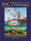
"The Tritones of Mass Spectrometry" Oil painting by Julie Newdoll, www.brushwithscience.com, 24" x 30", sold.
The Tritones are sea gods which normally do the bidding of Poseidon, king of the seas in Greek Mythology, but they have come to San Francisco for The 10th Annual Symposium on Mass Spectrometry. The merman is tossing up strands of jewels, which represent proteins, towards the surface of the bay. The mermaid holds a shell that allows a stream of water droplets through a hole in the center, alluding to the principal of mass spectrometry whereby a stream of water droplets containing the proteins of interest are sent into an electric field and become charged. Once the droplets containing protein "necklaces" reaches the sky, they are struck by a bolt of lightning by the helpful Zeus. This reflects the step in Mass Spectrometry where electricity is used to chemically break the protein into smaller, ionized pieces, which are sorted and analyzed according to their charge and weight. Strands of pearls attached to large gems represent epigenetic modifications to the proteins, a valuable piece of information gathered using this technique. Poster and program cover for the Tenth International Symposium on Mass Spectrometry in the Health and Life Sciences: Molecular and Cellular Proteomics, August 21-25, 2011.
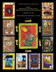
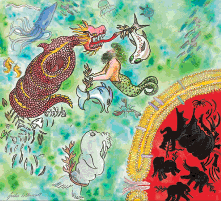
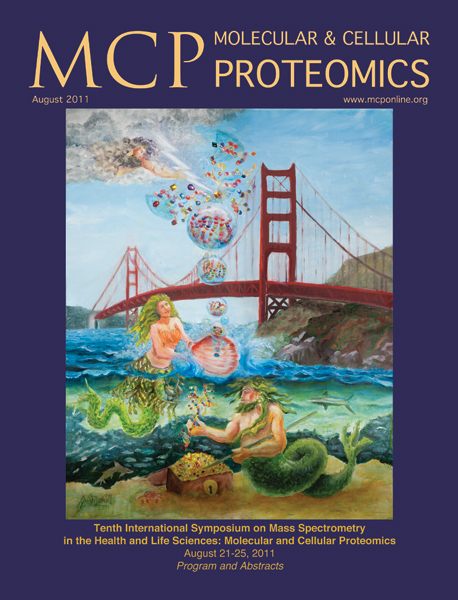
Digital collage of watercolor paintings for Cover of SUMO, Ubiquitin, UBL Conference Proceedings 2010 hosted by Dr. Edward T.H. Yeh
Cover of SUMO, Ubiquitin, UBL Conference Proceedings 2010, MD Anderson Cancer Center, organized by Dr. Edward Yeh
Ubiquitin-mediated proteasomal degradation (Center): The dragon (proteasome) breaks up a stretch of polypeptide, tagged for degradation with a string of gold medallions (ubiquitin) bearing the image of Alfred Nobel to symbolize the award of the Nobel Prize in 2004 to Avram Hershko, Aaron Ciechanover, and Irwin Rose. The center card is surrounded by keys that open up locks in the surrounding cards.
Starting from upper right in clockwise rotation: SUMO-mediated proteasomal degradation: Based on the oil painting SUMO and SENP1 wrestling over the Goddess of Hypoxia (HIF1a), 2007 The Goddess stands on a high mountain peak, where the air is thin (hypoxic), sending out newly formed blood vessels from her gown and blood cells from her sleeves. SUMO is attempting to grab the Goddess to throw her to the Dragon (proteasome), lurking below them in the clouds. SENP1 wields a sword trying to attack SUMO, separating him from the Goddess and saving her from the jaws of the dragon. Cell 131:584, 2007.
Immune response: The Inca rose from a waning tribe to an empire through "strong leadership, strategic alliances, and skill in warfare" (Gods of Order, Gods of War, A Comparison of the Major Gods and Myths of the Inca and Aztec, by Gini Graham Scott), in much the same way developing a good immune system boosted survival of organisms.
Neurological diseases: After the painting Nerve Eye on Channel Two, 40" x 40", 1992.
Cardiovascular diseases: Heart was rendered using watercolor.
Cell division: From Spindles of Necessity #3 (Anaphase) oil on canvas 3' x 4'.
Circadian rhythm: From “Dance of the Clock Gene Proteins”, . Cell 128: 59, 12 2007.
Signal transduction: From Cell Signals and Mayan Legends, 26" x 33", 2001. Growth hormone binds to the receptor tyrosine kinase (blue house structure) which attracts the protein grb2 (grandmother). This triggers a series of events which activate the proteins necessary for cell division.
DNA repair: Isis is an ancient Egyptian mother goddess associated, among other things, with healing.
DNA replication: Stringing beads is an ancient past-time. Egyptians were stringing nucleic acid and phosphate backbone beads during DNA replication.
Cancer: Rendering of cancer cell after image courtesy of Zena Werb, UCSF.
All artwork by Julie Newdoll. Full credits can be found in www.brushwithscience.com
SUMO and SENP1 wrestling over the Goddess of Hypoxia (HIF1alpha). Painting by Julie Newdoll, based on Cell article 131:584, 2007, sold.
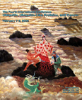
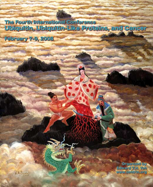
Cover of the Fourth International Conference on Ubiquitin, Ubiquitin-Like Proteins, and Cancer. February 2008.
This piece was a special commission by Dr. Edward T.H. Yeh. It is based on the paper cited above (Cell article 131:584, 2007), "SUMO-specific protease 1 is essential for stabilization of HIF1alpha during hypoxia." Cheng J, Kang X, Zhang S, Yeh ET.HIF1alpha stands on a high mountain peak, where the air is thin (hypoxic), sending out newly formed blood vessels from her gown and blood cells from her sleeves. SUMO is attempting to grab the Goddess to throw her to the Dragon (proteasome), lurking below them in the clouds. SENP1 wields a sword trying to attack SUMO, separating him from the Goddess and saving her from the jaws of the dragon.

Cells Both Dead and Alive as Calabash Fruit in the Tree Where Blood Moon is Impregnated by One Hunahpu, 33" x 44", 2000, sold. Prints available.
This painting was originally a special commission for Dr. Zbigniew Darzynkiewicz. The premise for this painting resides in the paradox of being both dead and alive at the same time. In the tree, one can find “Schrodinger’s cat”, in reference to the analogy used in quantum physics where a cat can be both dead and alive in the quantum world. The large red fruits in the tree are calabash fruits, modeled after cells which have been studied both while dead and alive. The large, full fruits are like healthy living cells and the multi-lobed fruits are more like apoptotic cells, or cells that died via a preprogrammed internal mechanism for cell suicide*. The woman sitting in the moon is Blood Moon, from an ancient Mayan legend found in the Popul Vuh**. She holds out her hand to accept a fruit from the tree. The central fruit with a skull imbedded in it is the head of One Hunahpu. He has been beheaded and his head stuck in a tree by the lords of the underworld after going there to accept their challenge to a ball game. Instead of giving her one of the fruits, he spits in her hand, saying,
“It is just a sign I have given you, my saliva, my spittle. This, my head, has nothing on it--just bone, nothing of meat. It’s just the same with the head of a great lord: it’s just the flesh that makes his face look good. And when he dies, people get frightened by his bones. After that, his son is like his saliva, his spittle, in his being, whether it be the son of a lord or the son of a craftsman, an orator. The father does not disappear, but goes on being fulfilled. Neither dimmed nor destroyed is the face of a lord, a warrior, craftsman, orator. Rather, he will leave his daughters and sons. So it is that I have done likewise through you. Now go up there on the face of the earth; you will not die.”
Inspired by the paper "The Schrodinger's Cat Quandary in Cell Biology: Integration of Live Cell Functional Assays with Measurements of Fixed Cells in analysis of Apoptosis", by Xun Li and Zbigniew Darzynkiewicz, Experimental Cell Research 249, 404-412, 1999.
Translation of the "Popul Vuh: The Mayan Book of the Dawn of Life", by Dennis Tedlock, Simon & Schuster Trade, 1995, ISBN: 0684818450
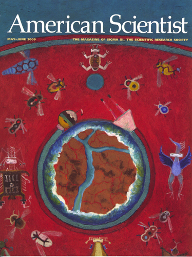
The First World was Red 30" x 40" Oil / mixed media on canvas, painting for sale $3,000.
On the Cover of American Scientist, May-June 2009.
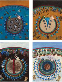
In her five-painting series Emergence, Julie Newdoll relates the five worlds of the Diné (Navajo) creation story to current scientific thinking on the origin of life. The cover painting, The First World Was Red, depicts a Hadean world in which primordial molecules are represented as Air Spirit People. Following (above) are worlds of blue, yellow, black-and-white, and Island World, eras linked by the artist to sugar, polymers, RNA, and finally, the emergence of the living cell. In "The Origin of Life," James Trefil, Harold Morowitz and Eric Smith present a new approach to the origin of life called Metabolism First: An ancient cycle of reactions still to be found in the core of modern metabolism reproduced its own constituents via reaction network that could then be acted upon by chemical selection, leading to increased complexity and finally life. Cover art courtesy of Julie

"Dance of the Clock Gene Proteins", 36" x 42", 2007, sold.
The figures are inspired by vase paintings from ancient Greece, and seals from Sumer. They dance around a very old sundial inspired by the Chaldean hemicyclium. There are twelve dancers, who represent various proteins that become active at specific times of the day or night in order to regulate our circadian rhythm. The sky was divided into twelve parts during the time of Ahaz in Egypt when the Chaldeans were perfecting their sundial. This lead to the division of the day into twelve parts as well (Sundials; History, Theory and Practice by Rene' R.J. Rohr).This painting was a special commission by Louis Ptacek.
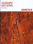

Dawn of the Double Helix, 18" x 25", 2003, sold.
Cover of Nature Reviews Genetics, July, 2003. This is the first painting in the series Life Forms, which explores the basic structures of our existence; the origin of ourselves. This painting uses figures to represent the structure of DNA, our genetic material which holds the information for what we look like and how we work. DNA is a double helix, where one strand is oriented in one direction and the other strand points in the opposite direction. Notice that on one side of the ladder of figures all the heads are looking upward, and on the other side their heads are all pointing downward. The male-female tension between the scientists involved in the discovery of the structure of DNA is another element to this work. This painting was completed in 2003, the year we celebrated the 50th anniversary of solving the structure of DNA. The human interactions in the painting symbolizes the ever changing give and take between the two strands of the helical structure and the molecules from which they are formed. The delicate bonds between some elements of the structure and strong bonds between others, their balance and cooperation, invokes a kind of dance of the molecules. *Inspired in part by the book, “Rosalind Franklin: Dark Lady of DNA” by Brenda Maddox, Harper Collins Publishers, 2002.

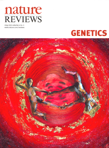
Base Pair, 18" x 24", oil and mixed media on canvas, 2003. Private Collection (sold). Prints available.
Cover of Nature Reviews Genetics, May, 2003. The dance and the tension between residues, people, male and female. Rosalind Franklin's famous X-ray diffraction photograph of DNA commands the center of the image.
The spiraling couple, frozen for an instant, is exposed to us, revealing the tension between the bases, between male and female, between the scientists involved in the discovery of the structure of DNA. The delicate hydrogen bonds between bases is the premise for a dance that can never stop as long as life continues.
Inspired in part by the book "Rosalind Franklin; The Dark Lady of DNA" by Brenda Maddox, Harper Collins Publishers, 2002.
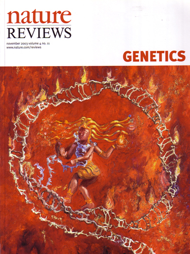
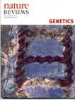
Shiva as Telomerase inside of a Telomere Loop, oil and mixed media on canvas, 2003, 32" x 38", Private Collection (sold). Prints available.
Cover of Nature Reviews Genetics, November, 2003. Shiva is the dancing manifestation of the Hindu god Nataraja. The dance of Shiva represents a delicate balance of creation and destruction, preservation and evolution, a life force as well as death. Here Shiva is dancing inside of a telomere loop, acting as the enzyme telomerase. The telomere is like the shoelace cap on the ends of a chromosome, keeping the lace (chromosome) from unraveling. Each time a human cell divides, all the chromosomes must also divide. The telomere gets shorter when the cell divides, and the protein “telomerase” inside the telomere binding complex lengthens the telomere again. Telomerase also stabilizes the telomere. The telomere has now been shown to be a large loop called a telomere loop next to a smaller loop, the D-loop.
Telomerase was once thought to be a potential key to the fountain of youth. If it were introduced into older people that no longer make telomerase in their cells, would their cells go on dividing? Cancer cells, on the other hand, have plenty of telomerase to perpetuate their eternally dividing cells. This is still a point of controversy, but it is certain that telomerase is an important enzyme, and one that is being studied carefully.
Inspired in part by the paper "Mammalian telomeres end in a large duplex loop." Griffith JD, Comeau L, Rosenfield S, Stansel RM, Bianchi A, Moss H, de Lange T. , Cell 1999 May 14;97(4):503-14. Information on Shiva from "Nataraja in Art, Thought and Literature" by C. Sivaramamurti, Thomas. Published in Nature Reviews Genetics 4, 845 (2003).
Cell Signals and Mayan Legends, oil on canvas, 26" x 33", 2001. Prints available. Available for purchase.
(The long title Mayan Hero Twins Hunahpu and Xbalnque as E2F and DP Receiving the Message to Begin Transcription for a New Cycle of Cell Division)
The first in a series, this painting shows the beginning of a cycle of cell division juxtaposed with a Mayan story about the hero twins Hunahpu and Xbalnque. Both stories have a similarly indirect way of getting their messages from one place to another.
In the legend, the hero twins are playing ball and are making such a racket, the underworld lords decide to summon them to the underworld and challenge them to a more dangerous game of ball with them. The send owls to deliver their message. This message goes from the owls to the house of their Grandmother. She gives the message to a louse. A toad eats the louse to carry the message faster. The toad is swallowed by a snake to bring the message even faster. Then a falcon eats the snake and carries the message via air to them. They see the falcon, shoot him in the eye to bring him down, and in return for fixing his eye he spits up the snake. The snake spits up the toad, who spits up the louse, who finally relays the message.
Before a cell will divide, a message must get to the nucleus of the cell. A growth hormone reaches the outside of the cell and binds to the receptor tyrosine kinase (blue house structure). This attracts the protein grb2 (grandmother) to the internal portion of the receptor. grb2 interacts with the protein sos (louse) which binds to the protein ras (toad). A kinase cascade is then triggered into action (snake) which ultimately causes the production of cyclin D/kinase in the nucleus of the cell (falcon). This causes the protein pRB (the ball) to be removed from the e2f/dp complex, which allows them to participate in transcription of the proteins necessary for cell division.
Tyrosine kinase receptor image courtesy of Moosa Mohammadi, NYU. E2F/DP consultation Jacqueline Lees. Ras and Sos consultation J. Kuriyan and L. Leighton. pRB image by permission "Structure of the retinoblastoma tumor-suppressor pocket domain, HPV E7" from Jie-Oh Lee, Alicia A. Russo and Nikola P. Pavletich, Nature. 1998 Feb 26;391(6670):859-65. 2erk from the protein databank , "Phosphorylated Map Kinase ERK2", B.J.Canagarajah ,E.J.Goldsmith. Cyclin A from 1fin from the protein databank, P.D.Jeffrey, A.A.Russo,N.P.Pavletich. RasMol used to create PDB protein images. Translation of the "Popul Vuh: The Mayan Book of the Dawn of Life", by Dennis Tedlock, Simon & Schuster Trade, 1995, ISBN: 0684818450
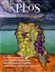

Cover of "Public Library of Science" Journal, December 2003, digital painting. Prints available The cover depicts the structure of the newly-discovered, water-selective aquaporin AqpZ. Like its relative in the "Ishtar. . ." painting above, this protein is a membrane protein. It controls the flow of water into a cell. Five water molecules are seen in the channel defining the conductance pathway. This painting is a bit of a departure from the other paintings displayed on the rest of this web site. Using the symbolism I have developed for my other paintings, I have placed a scientific representation of the AqpZ protein into a conceptual world. The protein is now in the same mythical surrounding which is depicted in the Ishtar painting above, but the rest of the Ishtar story has been replaced with an actual aquaporin structure. In addition to relating the new protein structure to its glycerol channel cousin, I am using the symbolic language of art to explore the properties of a cell and its surroundings; the calm internal blue-green of the membrane; the rough exterior surface of the membrane; the hot bed of activity inside of the cell and the approaching storm bringing water outside the cell; the ancient origins of our cellular systems.
Commission by Dr. Robert Stroud, UCSF. Journal article Architecture and Selectivity in Aquaporins: 2.5 Å X-Ray Structure of Aquaporin Z, David F. Savage, Pascal F. Egea, Yaneth Robles-Colmenares, Joseph D. O'Connell III, Robert M. Stroud. PLOS Volume 1, Issue 3, December 2003.
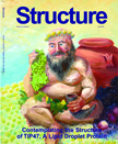

Cover of "Structure" Journal, July 2004, Private Collection; Prints available without text.
On the cover: The obese Greek god Silenus contemplating the crystal structure of the lipid droplet protein TIP47. TIP47 contains a hydrophobic pocket that is conserved in the adipocyte lipid droplet protein, perilipin. Perilipin and its hydrophobic pocket are potential targets for anti-obesity drug design.
Structure of a Lipid Droplet Protein: The PAT Family Member TIP47 Sabrina J. Hickenbottom 1, Alan R. Kimmel 1, ConstantineLondos 1, and James H. Hurley. Structure, Vol 12, 1199-1207, July 2004

Nanotechnology II, 3' x 4', 1992, private collection.
This DNA ladder is simultaneously creating itself at one end and destroying itself at the other end. A reflection of the need for creation, destruction, and disposal of nanotechnology devices in biological systems and experimental environments. See the "Nanotechnology I" description above for more information.

Nanotechnology I, 20" x 24", 1992, private collection.
Nanotechnology is the science of manipulating atoms to build tiny machines. These machines could be computers, chemical factories, or other types of mechanical device. It has been proposed that DNA be used as a scaffold to put tiny molecular machines onto. It has been shown that DNA can be used to form ladders and cube structures. These would make stable structures to attach tiny atomic sized machines to.
Inspired in part by the paper "Synthesis from DNA of a molecule with the connectivity of a cube", Junghuel Chen and Nadrian C. Seeman, Letters to Nature, Vol. 350, 1991.

Nanotechnology III, 20" x 24", 1992. Private collection.
A possible result obtained after folding DNA into a ladder shape. A solid scaffolding onto which tiny molecular machines could be attached and made to work together. See the "Nanotechnology I" description above for more information.

FAS Sisters Receiving a Death Signal, 25" x 33", 2000. Private Collection. Prints available.
Three Fas identical sister proteins protrude above the cell surface. They are being organized by a death signal in the form of three Ravens, which represent the homotrimeric molecule, CD95L. Once the external trimeric FAS molecule has bound to its ligand, CD95L, the pathways of apoptosis are triggered inside the cell.
Inspired in part by the paper "Death Receptors: Signaling and modulation", by Avi Ashkenazi and Vishva M. Dixit, Science, Vol. 281, 1305-1308, 1998, and the paper "BCL-2 family members and the mitochondria in apoptosis", Aton Gross, James McDonnell, Stanley Korsmeyer, Genes and Development, 13:1899-911, 1999.

The transportation of Ra through a Vault Protein out of the Night Sky and into the Birth of the Sunrise, 33" x 44", 2001. Private Collection (sold). Prints available.
In the ancient Egyptian legend, “The Regions of Night and Thick Darkness”, Ra, the sun king, dies each night and makes a journey in his “Boat of Millions of Years” through the otherworld of the night sky. After traveling through twelve regions of the underworld country, Duat, each guarded by a gate and a terrifying snake, he is once again reunited with his soul in the form of the scarab beetle, Khepera. The sun king is thus alive again, and a new sunrise is born.
This mysterious process of daily transport to create the cycle of night and day is shown here inside of a capsule-like particle composed of proteins called “the vault”. The function of the vault particle is still a scientific mystery, although there is some evidence that it is involved in cellular transport. The structure of the vault is a “hollow, barrel-like structure with two protruding caps and an invaginated waist”. In this painting we are inside of a vault particle looking out through one of its end caps. At the caps of the structure there are 12 RNA strands of a very special type, similar to the telomeres at the endcaps of chromosomes (pieces of DNA which help protect the chromosome and keep the ends from fraying). The night sky was considered a vault by ancient peoples that did not realize the earth was round.
This story may be found in “Legends of Ancient Egypt”, by M.A.Murray, Dover Publications, Inc., 2000, ISBN 0-486-41137-0. Inspired by the papers “Structure of the vault, a ubiquitous cellular component”, Lawrence B Kong , Amara C Siva , Leonard H Rome and Phoebe L Stewart , Structure 1999, Vol 7 No 4; and “RNA location and modeling of a WD40 repeat domain within the vault”, Lawrence B. Kong, Amara C. Siva, Valerie A. Kickhoefer, Leonard H. Rome, and Phoebe L. Stewart , RNA(2000), 6:890–900+
Woman Contemplating the Increasing Speed of her Biological Clock: 33" x 44", 1999, Private Collection.
A woman is born with thousands of eggs (oocytes) at birth. During her lifetime, she will ovulate only about 400 eggs. The rest of these eggs seem to self-destruct via the mechanism of apoptosis, or programmed cell death. What causes these eggs to die? If they died at a constant rate until menopause, a woman would be losing two eggs a minute. It seems, however, that we lose fewer eggs during the early part of our life, and this egg loss gets faster and faster as a woman ages. The oocyte in the background is like a waning moon. Egg loss has reached the tenth hour of her biological clock.
Inspired in part by the paper "Oocyte Apoptosis: Like Sand through an Hourglass", Yutaka Morita and Jonathan Tilly, Developmental Biology 213, 1-17, 1999. Figure originally in "Prolongation of ovarian lifespan..." by Perez et. al, Nature Genetics. vol21 #2, 1999. Special thanks to Chris Gralapp.
Figures in a Beta Sheet, oil and mixed media on canvas, 18" x 24", 2003. Private Collection (sold). Prints available.
Human figures arranged in a beta sheet, a structure found in folded proteins. The flowers are like the sidechains each amino acid holds out.
Inspired by the beta sheet structure found in proteins, discovered by Linus Pauling and Robert Corey, and Herman Branson.

RNA as the New Dogma, oil and mixed media on canvas, 24" x 33", 2003, sold.
Prints available. Purchase Info. Human figures are assembled here into a possible RNA shape. DNA is often thought of as our primary life giving molecule. We may have evolved from RNA in a form that was capable of self replication. Scientists are still discovering more that RNA is responsible for in our cells. Although the bases that make up RNA are very similar to DNA, they can create structures very different from the double helix that we know DNA to typically, but not always, form.
RNA does much more than previously thought, according to John Mattick of the University of Queensland. His research and that of his collaborators may overturn the Central Dogma, the current thinking on how we control and express our genes, in which RNA plays mainly a messenger role. RNA may do much, much more. This is a topic for a new painting series to come.
The figures here are more ancient and ambiguous than those in "Dawn of the Double Helix". Surrounded by forces without and within, these figures collect around pearl-like ions, and brush against metallic flakes of gold. Surfaces such as iron pyrite (fool's gold) may have served as substrates for the first self replicating molecules to form upon.


The Descent of Ishtar into the Underworld through the Ancient Gates of an Aquaporin Glycerol Channel33” x 44”, 2000. Private Collection. Prints available.
Ishtar was a goddess in the times when dreams were just as important as the waking reality; prophetic and real. Her story is revealed to us on clay tablets(1), the earliest version from around 1,600 B.C, unearthed in modern day Iraq. Written in cuniform, the oldest known written language, her ancient story presented itself to me during my search for an analogy between a most ancient biological gate and a mythological counterpart. This biological gate is found in membrane proteins that allow only water or glycerol to cross the membrane and enter a cell, and has been conserved for over four billion years.
A small handful of amino acids compose this gatekeeping system, and every species which contains a protein for conducting water or glycerol into a cell also contains this gateway package of amino acids. The first structure to be determined for this kind of channel was for a protein called the GLPF receptor (glycerol facilitator protein)(2). This protein is represented here, where the pillars of the channel in the painting mimic the arrangement of the seven helices which compose the GLPF receptor, with two of the helices stripped away to reveal the inside of the channel. The stairway portrays the connections between the helices that allow specific molecules to enter the channel, and the ancient gate is shown in its amino acid structure on the stairway.
In the Ishtar story, her descent into the underworld is strikingly enzymatic. She must pass through seven doors, losing a piece of clothing at each door, until she arrives naked into the underworld. Glycerol is like food for a bacteria, and once it passes through the gates of the GLPF receptor, it enters the cell to be broken down and digested into its various components. Ishtar descends the staircase and passes through the gates to enter the underworld using the same route a glycerol molecule would travel through this structure.
(1) “Myths from Mesopotamia, Creation, The Flood, Gilgamesh and Others”, by Stephanie Dalley, 1989, Oxford University Press, ISBN 0-19-283589-0 (2) “Structure of a glycerol-conducting channel and the basis for its selectivity”, Science, 2000 Oct 20;290(5491):481-6 . Fu D, Libson A, Miercke LJ, Weitzman C, Nollert P, Krucinski J, Stroud RM.
Zoka Coffee

Zoka Coffee Dynamic Drive

Dynamic Drive