
...Julie Newdoll merges life science and culture, myths and molecules in her
paintings, music, journal covers and science games.
Shakespeare:
A Mirror up to Science
 Microscopy
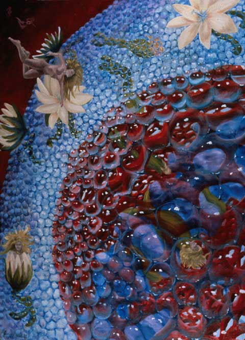
30" x 40", 1991, private collection. Prints available $450.
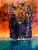
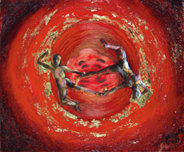
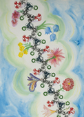
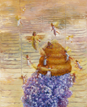
The cycle of estrogen takes this small molecule through the bloodstream until it reaches cells such as uterine cells. An estrogen molecule has just transformed into the floating figure. She is attracted to the lotus flowers, which represent estrogen receptors on the surface of a cell. The flower captures her, and together they enter the cell and then the red cell nucleus. They then are bound to the purple DNA inside the nucleus. At this point, they cause the DNA to ultimately produce substances that cause the feelings and actions that estrogen is meant to trigger. Estrogen causes many, many things to occur.
Reference for description, "Biochemistry", Lubert Stryer, p. 1001, 1988.ISBN 0-7167-1843-X.
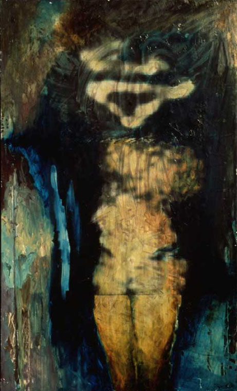
Conception, 30" x 40", 1990, private collection.
This painting was inspired by an electron microscope image of a drosophila sperm fertilizing an egg. Microscope image taken and processed in the labs of Professor John Sedat and Professor David Agard, University of California at San Francisco.
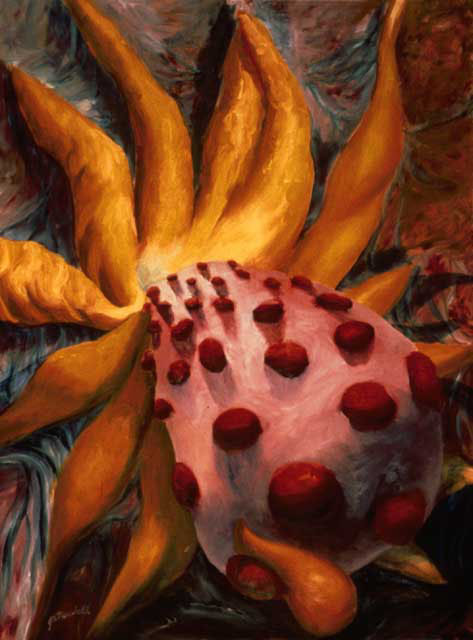
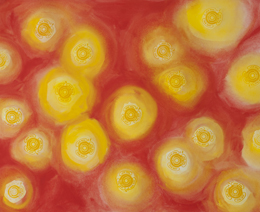
Ovariation, oil on canvas, 30" x 40", 1990, private collection.
This painting was inspired by an image of a drosophila ovary releasing lots of eggs on its surface. From here, it became a human ovary. In humans, there is a large searching tentacle, called a fimbriae, which sweeps over an ovary searching for an ovulated egg. Once it finds the egg, it draws it into the fallopian tube. This egg has released an abnormal amount of eggs. Microscope image taken and processed in the labs of Professor John Sedat and Professor David Agard, University of California at San Francisco.
![]()
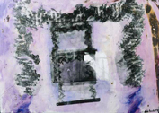
Nuclear Window, 30" x 40", 1990, In the collection of Dr. Dan Friend. Prints available $450.
The nucleus of a cell is made of a network of proteins. This painting was inspired by an image of one such protein, stained and viewed through a microscope. The pores may be places where other proteins attach. The protein may be a structural one that keeps the nucleus a more stable spherical shape.
Inspirational image from Interphase nuclear envelope lamins form a discontinuous network that interacts with only a fraction of the chromatin in the nuclear periphery., Paddy MR; Belmont AS; Saumweber H; Agard DA; Sedat JW, Cell 1990 Jul 13;62(1):89-106.
Notch Gene Figure Head, 3.5' x 5', 1993, Not For Sale.
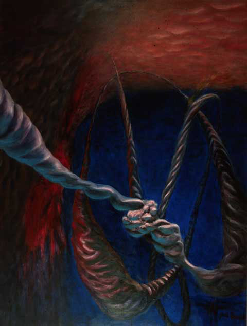
This painting began as a figure from a research paper, which looked very much like a human female figure to me. The triangular "head" is the the "notch gene" stained and photographed under a microscope. The body is the microscope image of the dna. The notch gene is found at the pelvic area of the figure. The legs of the figure were added as part of the painting.
Inspired by a figure in the paper, Precise determination of the molecular limits of a polytene chromosome band: regulatory sequences for the Notch gene are in the interband, Rykowski MC; Parmelee SJ; Agard DA; Sedat JW, Cell 1988 Aug 12;54(4):461-72


Programmed
Cell Death:
Apoptosis