

 View Portfolio |
 Inside Time |
 Shows |
 About/Contact the Artist |
Early Works
These paintings are the earlies works featured on these pages. A variety of microscope images were key inspirations for many of these paintings.
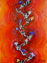
Plight of the Estrogen Ladies,
30" x 40", 1991, private collection.
The cycle of estrogen takes this small molecule through the bloodstream until it reaches cells such as uterine cells. An estrogen molecule has just transformed into the floating figure. She is attracted to the lotus flowers, which represent estrogen receptors on the surface of a cell. The flower captures her, and together they enter the cell and then the red cell nucleus. They then are bound to the purple DNA inside the nucleus. At this point, they cause the DNA to ultimately produce substances that cause the feelings and actions that estrogen is meant to trigger. Estrogen causes many, many things to occur.
For description, "Biochemistry", Lubert Stryer, p. 1001, 1988.ISBN 0-7167-1843-X
)
Conception, 30" x 40", 1990, private collection.
)
Nerve Eye on Channel Two, 40" x 40", 1992, private collection.
Nerve eye on channel two is a nerve ion channel, too. The neurons in our brain have many ion channels, which control the firing of neurons. An ion channel is a barrel shaped protein that allows small molecules, ions, to pass from outside of a cell to inside a cell, or vice versa. The brain contemplates this phenomenon.
Computer generated Ion channel model by Robert M. Stroud and Julie Newdoll.
)
Evolution of Sight, 30" x 40", 1991, private collection.
There is a type of bacteria, Cyanobacteria, that uses light to produce energy. On its surface, there is a field of proteins that can capture light. This protein is probably related to the same types of proteins which are in our eyes. Bacteria use a protein called "Bacteriorhodopsin", and humans have a protein called "rhodopsin", and they are very similar in structure.
Special thanks to George Turner for consultation on this painting.
)
Ovariation, 30" x 40", 1990, private collection.
This painting was inspired by an image of a drosophila ovary releasing lots of eggs on its surface. From here, it became a human ovary. In humans, there is a large searching tentacle, called a fimbriae, which sweeps over an ovary searching for an ovulated egg. Once it finds the egg, it draws it into the fallopian tube. This egg has released an abnormal amount of eggs.
)
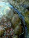
Nuclear Window, 40" x 30", 1990, private collection.
The nucleus of a cell is made of a network of proteins. This painting was inspired by an image of one such protein, stained and viewed through a microscope. The pores may be places where other proteins attach. The protein may be a structural one that keeps the nucleus a more stable spherical shape.
Inspirational image from "Interphase nuclear envelope lamins form a discontinuous network that interacts with only a fraction of the chromatin in the nuclear periphery.", Paddy MR; Belmont AS; Saumweber H; Agard DA; Sedat JW, Cell 1990 Jul 13;62(1):89-106.
Knowledge, 38" x 38", 1991, private collection
Electronic stamping phase.
Special thanks to my father for consultation.
)